New radioactive agents for imaging of neuroprotective immune cells in MS
23.03.2022Scientists in Amsterdam are working on developing new radioactive agents that can be used to the imaging of the protective state of the brain-specific immune cells in MS called microglia.
The role of microglia in multiple sclerosis
The brain has its own specific immune cells called microglia. Upon inflammation in the brain, these immune cells become activated and play a key role in multiple sclerosis (MS) related disease activity. The activation of microglia is very complex and covers a wide range of activation states.
Depending on their activation state evidence suggests that these cells can have both a toxic function, as well as a protective and repairing function. Within the context of MS, the toxic microglia drives inflammation and is responsible for disease progression. In contrast, the protective microglia are involved in repairing damaged nerve tissue and restraining the inflammatory responses.
Several therapies that are currently in development focus on forcing microglia into the protective state, thereby preserving brain tissue, halting disease progression, and thus helping patients. Despite lots of progress, there are still many unanswered questions about microglia. To shed light on its function and to fill the current knowledge gaps, we need to study these cells at a molecular level.
How can we study the different types of microglia up close in the living brain?
Positron Emission Tomography (PET) is a non-invasive imaging technique that makes use of a radioactive agent to study a biochemical process. By injecting a very little amount of these radioactive agents into a human patient, a PET camera can detect the amount of radioactivity in each organ without the need of surgery.
For investigating the changes between toxic and protective microglia, two radioactive agents are needed. One agent should selectively bind to toxic microglia and the other should only bind to the protective state of microglia. More specifically, each one of the radioactive agents should bind to a protein over-expressed on the particular state of microglia that are being studied.
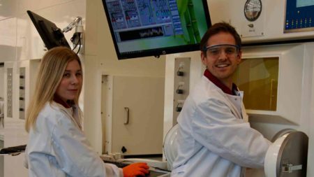
The protein, one for each microglia state, can be seen as an indicator for that specific microglia. Johanna Stéen and Berend van der Wildt have each received a personal research grant to work on developing these types of radioactive agents to study microglia. The funding was obtained from the European Union’s Horizon 2020 research and innovation programme and from the Dutch research foundation NWO.
The research is carried out at the Department of Radiology & Nuclear Medicine, Amsterdam UMC and the Division of Medicinal Chemistry at the Vrije Universiteit in the research groups of Bert Windhorst and Iwan de Esch.
An innovative approach starting at the basis of disease
The research group has already developed an agent to study the toxic state of microglia. At the moment this agent is used in exploratory studies in MS patients. The focus is now to develop a radioactive agent that can bind to the P2Y12 receptor, an protein indicative for the protective microglia.
Several radioactive agents have been designed and tested in animal models and more are in development. Obtaining sufficient brain uptake is one of the many challenges with developing this type of agent. To be able to bind to the P2Y12 receptor in the brain the radioactive agent has to cross the blood-brain barrier, which is a protective layer that prevents molecules from entering the brain.
To minimize the risk of developing an agent that in the end cannot enter the brain, the team use computational methods to design agents that not only bind extremely well to the P2Y12 receptor, but also have good properties for crossing the BBB.
Does this mean new drugs for MS?
With a successful radioactive agent targeting the P2Y12 receptor, the changes in microglia activity can be monitored and measured at different stages of MS . This insight can pave the way for improved treatment monitoring and diagnosis, as well as boosting drug development to bring us yet one step closer to treatments that actually halt MS progression.
For more info:
https://cordis.europa.eu/project/id/892572
https://www.amsterdamumc.org/en/research/institutes/amsterdam-neuroscience.htm
 Your Account


